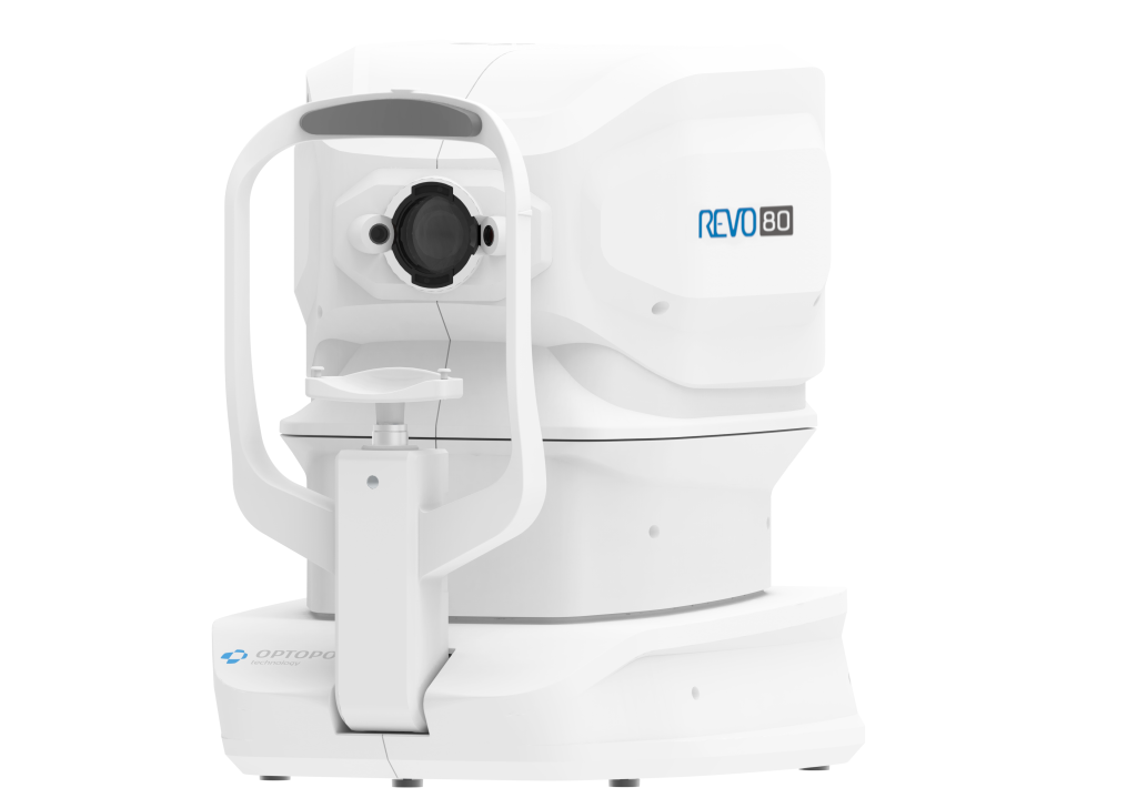
Optopol REVO 80 OCT

Product Ref: OPTO-REVO-80
Download Product BrochureAdd this item to your wishlist by clicking below
The Optopol REVO 80 provides 80,000 A scans per second. It joins the REVO range sitting in-between the REVO 60 and REVO FC
The REVO 80 facilitates the use of the optional Biometry,Topography and Angiography software as the scan time is faster and brings benefits for both clinicians and patients by reducing errors often caused by involuntary eye movements. The higher sensitivity spectrometer allows improved visualisation for finer detail.
RETINA
| Single 3D Retina examination is enough to perform both Retina and Glaucoma analysis based on retinal scans. | Software automatically recognize 8 retina layers. Thus allowing a more precise diagnosis and mapping any changes in the patient’s retina condition. |
GLAUCOMA
| Comprehensive glaucoma analysis tools for Quantification of Optic Nerve Head, Retina Nerve Fiber Layer, DDLS, Ganglion layer and Asymmetry | |
ANTERIOR
| For standard examinations no additional lens is required. Additional adapter provided with the device allows to make wide scans of anterior segment. |
FOLLOW UP
| High density of standard 3D scan allows to precisely track the disease progress. Operator can analyze changes is morphology, quantified progression maps or evaluate the progression trends. |
|
NETWORKING
A proficient networking solution increases productivity and an enhanced patient experience. It allows you to view and manipulate multiple examinations from review stations in your practice. Effortlessly helping to facilitate patient education by allowing you to interactively show examination results to patients. Every practice will have differing requirements which we can provide by tailoring a bespoke service. There is no additional charge for the server module.
Optional extras:
Biometry software
Topography software
Angiography software
Hewlett Packard/Bang & Olufsen 27inch embedded touch screen PC
A variety of table models to suit your exacting requirements
Please ask for further information.
TECHNICAL DATA
| Technology | Spectral Domain OCT |
| Light Source | SLED, Wavelength 840nm |
| Bandwidth | 50 nm half bandwidth |
| Scanning speed | 80,000 A-scan per second |
| Axial resolution | 5 µm in tissue |
| Transverse Resolution | 12 µm, typical 18 µm |
| Overall scan depth | 2.4 mm |
| Scan range | 3 to 12 mm |
| Scan types | 3D, Radial, B-scan, Raster, Cross |
| Fundus image | Live Fundus Reconstruction |
| Alignment method | Fully automatic, Semi-automatic |
| Retina analysis | Retina thickness, Inner retinal thickness, Outer retinal thickness, RNFL+GCL+IPL thickness, GCL+IPL thickness, RNFL thickness, RPE deformation, IS/OS thickness |
| Glaucoma analysis | RNFL, ONH morphology, DDLS, Ganglion analysis as RNFL+GCL+IP and GCL+IPL, OU and Hemisphere asymmetry |
| Anterior | Pachymetry, LASIK flap, Angle Assessment, AIOP, AOD 500/750, TISA 500/750 |
| Anterior Wide Scan | Angle to Angle view, Adapter required |
| Min. pupil size | 3 mm |
| Focus adjustment range | -25D to +25D |
| Dimension/weight | 382 (W) x 549 (D) × 462 (H) mm |
| Weight | 23 kg |
| Fixation target | OLED display (The target shape and position can be changed), External fixation arm |
| Power supply | 110-230 V, 60/50 Hz |
| Power consumption | 115 – 140 VA |
Accessories and other items can also be purchased by phone if you prefer. To make a telephone order, or to discuss any item purchase please call 01438 740823.

