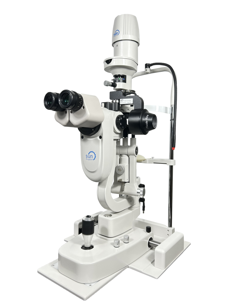
SK-MED SL-5C LED Slit Lamp with DA-1 Dry Eye Analyser

Web Price | £8,995 EX VAT |
Please click on the button below, to request a price.
Add this item to your wishlist by clicking below
Interested in this item? Would you like more information? Looking to buy more than one item?
Call our Sales department on 01438 740823
Combine and record all your anterior imaging in one instrument
SK-MED SL-5C
Proven Haag Streit design
5 mag options up to 40x
20 Million pixels Camera (The highest of any in -line slit lamp camera). High depth of field supplemented by front diaphragm controller
Coaxial back ground light -variable
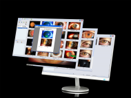
Imaging and archiving software included
Wide slit width – 0 to 14mm
Wide slit height – 1 to 14mm
Step-less variable LED light source
DA-1 Dry Eye Analyser
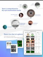
Fully comprehensive dry eye analysis
A comprehensive and professional dry eye examination and diagnosis program providing seven methods of dry eye detection functions to provide a reliable basis for clinical diagnosis
Multifunction – A full set of dry eye examinations can be completed in 7 minutes, effectively improving outpatient efficiency
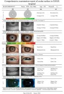
Detailed Dry Eye Test Report – The test report provides images and texts , the test results are clear and detailed helping doctors in recommending appropriate treatment
Non-invasive – Non-invasive inspection method, safer operation, improved patient comfort and promoting better patient cooperation
Automatic – Fully automated software analysis. Quantification of testing standards, for easier diagnosis by doctors
Convenient – Patient management system, which continuously records and tracks the development of dry eye conditions
Accurate Grading – Internationally recognized grading scales
Main functions of dry eye module
Imaging Analysis – Automatically analyzes the deficiency rate and categorizes results for determining fast-track patient treatment pathways
Gland Opening – HD eyelid image
Ocular Redness analysis – HD eye surface image, automatically recognize blood vessels, automatic ocular redness analysis scoring
Meibomian Glands Imaging analysis -Acinar-level HD image
Corneal Staining Analysis – Professional fluorescent corneal stain image, automatic analysis
Lipid Layer analysis – Uses a constant and uniform light source for detection. Provides the highest, lowest and average thickness values and provide automatic analysis
TMH – Optional infrared light/ visible light measurement. Can provide central average TMH
NIBUT – Provide first, average break-up time. Automatic analysis
PC Requirement
CPU, Model:intel i5, Speed:≥2.9 GHz, RAM ≥16G, Graphics card – Chips: nVidia GTX1650, Type: Discrete Graphics Cards -Display card capacity:4G -Hard Disk ≥1T – System Windows 10 64bit or Windows 11 64bit -Hard Disk Partition C: ≥100G D: ≥700G (At least two zones) – USB connector port USB 2 x 2 and 3 x 1 – Display Screen Resolution:1920*1080 – Laptops or all-in-one computers need to disable the camera that comes with the computer – When running dry eye programs, please close the anti-virus software – Turn off system updates – It is recommended to turn off the firewall
Accessories and other items can also be purchased by phone if you prefer. To make a telephone order, or to discuss any item purchase please call 01438 740823.


